Multi-modality Platform for Specific Imaging in Cardiology
MUSIC (Multi-modality Platform for Specific Imaging in Cardiology) is a software developed by the ‘Multimodal Data Science’ team of the IHU Liryc and the Inria Epione and Inria Carmen teams. The objective is to offer a large range of functionalities and processing chains dedicated to cardiology, both for diagnosis and for therapeutic guidance. MUSIC is made of several modules, such as the MUSICardio software, based on the medInria open source software. It offers :
About the MUSICardio tool
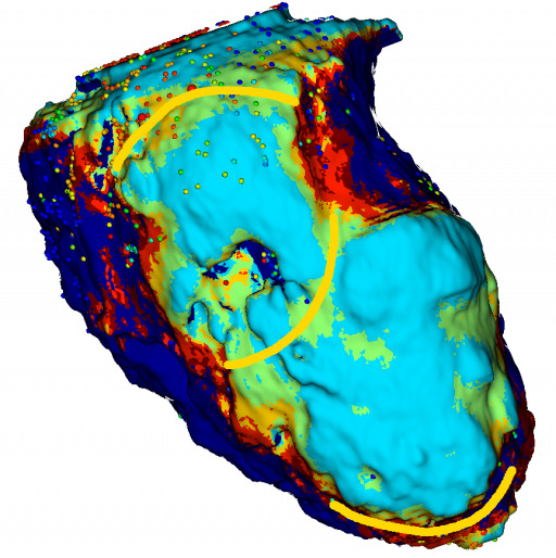
Segmentation, visualisation, filters, histogram, reformatting, registration and mesh management tools are proposed and allow the community to use an ergonomic and more efficient software system. Many imaging modalities are supported: MDCT, MRI, PET, VTK meshes, etc. In addition, the generated models are exportable to electro-anatomical 3D mapping systems used by cardiologists during interventions on cardiac rhythm disorders (catheter ablation).
MUSICardio offers image processing algorithms for diagnostic or prognostic purposes, processing chains intended to guide interventions on atrial and ventricular arrhythmias through imaging. Thus, MUSICardio has been used to guide ventricular tachycardia (VT) ablations in more than 300 procedures in our centre, and more than 1500 in the international MUSIC network.
MUSICardio is a software platform that allows multi-parametric data from the various IHU teams (structural, mechanical, haemodynamic, electrical, etc.) to be analysed in the same environment, and to interface with simulation software platforms such as SOFA, CEPS and CARP in order to develop modelling/simulation strategies tailored to the patient.
An international consortium of centres specialising in TV was created within the framework of the MUSIC project. It now includes more than 30 international centres. Participating centres send their anonymised data to our team through a web portal. Our radio handlers process them within hours and send them back to the partner centre in time for the intervention. Every day, cardiologists from all over the world use this technology to guide their interventions and visualise their catheters in real time within a personalised virtual heart produced in Bordeaux. In addition, this multicentre network allows the creation of an international registry of structural cardiac pathologies including imaging and electrophysiology data.
About the team
Our team: The MUSIC project was developed by a multidisciplinary team including doctors, researchers, computer scientists, and radiographers.
Project contributors :
Responsible for development :
- Florent Collot (Developer)
- Mathilde Merle (Developer)
Developers :
- Loic Cadour (Developer)
- Julien Castelneau (Developer)
- Nordine El Baraka (Developer)
- Mehdi Juhoor (Developer)
- Pauline Migerditichan (Developer)
- Florian Vichot (Developer)
PhD student :
- Jatin Relan (PhD Student)
Scientific coordinator :
- Maxime Sermesant (Researcher)
Clinical coordinators:
- Hubert Cochet (Radiologist): cardiac image analysis
- Pierre Jais (Cardiologist): procedure guidance
Data analysis :
- Olivier Baris (Radio operator)
- Ludovic Germain (Radio technician)
- Bruno Soré (Radio manipulator)
- Jean Michel Thomas (Radio technician)
- Imaging team leader:
- Bruno Quesson (Researcher)
Team leader “Signal processing” :
- Rémi Dubois (Researcher)
Modelling” team, Inria “Carmen” team:
- Nejib Zemzemi (Researcher)
- Yves Coudière (Researcher)
To contact us: here
Latest news from the MUSIC project
Find out more about the MUSIC project from Cyril:
The MUSIC project detailed in the article “MUSIC: Cardiac Imaging, Modelling and Visualisation Software for Diagnosis and Therapy“
Watch the EquipEx Music presentation:
Click here to discover an article on MusiCardio, the digital simulation software for cardiology.
Video clip “The rhythm of the heart” (video)
Scientific publications
- Substrate Modification Using Stereotactic Radioablation to Treat Refractory Ventricular Tachycardia in Patients With Ischemic Cardiomyopathy. Qian PC, Quadros K, Aguilar M, Wei C, Boeck M, Bredfeldt J, Cochet H, Blankstein R, Mak R, Sauer WH, Tedrow UB, Zei PC.JACC Clin Electrophysiol. 2021 Jul 27:S2405-500X(21)00599-5. doi: 10.1016/j.jacep.2021.06.016.
- Pulsed field ablation prevents chronic atrial fibrotic changes and restrictive mechanics after catheter ablation for atrial fibrillation. Nakatani Y, Sridi-Cheniti S, Cheniti G, Ramirez FD, Goujeau C, André C, Nakashima T, Eggert C, Schneider C, Viswanathan R, Krisai P, Takagi T, Kamakura T, Vlachos K, Derval N, Duchateau J, Pambrun T, Chauvel R, Reddy VY, Montaudon M, Laurent F, Sacher F, Hocini M, Haïssaguerre M, Jaïs P, Cochet H.Europace. 2021 Jul 8:euab155. doi: 10.1093/europace/euab155.
- Cardiac Magnetic Resonance Imaging and Ventricular Tachycardias Involving the Sinuses of Valsalva in Patients With Nonischemic Cardiomyopathy. Ghannam M, Liang JJ, Attili A, Cochet H, Jais P, Latchamsetty R, Jongnarangsin K, Morady F, Bogun F.JACC Clin Electrophysiol. 2021 Jun 22:S2405-500X(21)00282-6. doi: 10.1016/j.jacep.2021.03.017.
- Role of endocardial ablation in eliminating an epicardial arrhythmogenic substrate in patients with Brugada syndrome. Kamakura T, Cochet H, Juhoor M, Nakatani Y, Ramirez FD, André C, Nakashima T, Krisai P, Takagi T, Tixier R, Chauvel R, Cheniti G, Duchateau J, Pambrun T, Derval N, Kusano K, Sacher F, Jaïs P, Haïssaguerre M, Hocini M.Heart Rhythm. 2021 Oct;18(10):1673-1681. doi: 10.1016/j.hrthm.2021.06.1188.
- Deep learning formulation of electrocardiographic imaging integrating image and signal information with data-driven regularization. Bacoyannis T, Ly B, Cedilnik N, Cochet H, Sermesant M.Europace. 2021 Mar 4;23(23 Suppl 1):i55-i62. doi: 10.1093/europace/euaa391.
- The value of cardiac magnetic resonance imaging and programmed ventricular stimulation in patients with ventricular noncompaction and ventricular arrhythmias. Gunda S, Ghannam M, Liang JJ, Attili A, Sharaf Dabbagh G, Cochet H, Lathkar-Pradhan S, Latchamsetty R, Jongnarangsin K, Morady F, Bogun F.J Cardiovasc Electrophysiol. 2021 Mar;32(3):745-754. doi: 10.1111/jce.14884.
- Impact of Intramural Scar on Mapping and Ablation of Premature Ventricular Complexes. Ghannam M, Liang JJ, Dabbagh GS, Siontis KC, Attili A, Cochet H, Jais P, Juhoor M, Latchamsetty R, Jongnarangsin K, Morady F, Bogun F.JACC Clin Electrophysiol. 2021 Jun;7(6):733-741. doi: 10.1016/j.jacep.2020.11.004.
- Magnetic Resonance Mapping of Catheter Ablation Lesions After Post-Infarction Ventricular Tachycardia Ablation. Dabbagh GS, Ghannam M, Siontis KC, Attili A, Cochet H, Jais P, Eng MJ, Latchamsetty R, Jongnarangsin K, Morady F, Bogun F.JACC Cardiovasc Imaging. 2021 Mar;14(3):588-598. doi: 10.1016/j.jcmg.2020.08.041.
- Clinical significance of myocardial scar in patients with frequent premature ventricular complexes undergoing catheter ablation. Ghannam M, Yokokawa M, Liang JJ, Cochet H, Jais P, Dabagh GS, Latchamsetty R, Jongnarangsin K, Morady F, Bogun F.Heart Rhythm. 2021 Jan;18(1):20-26. doi: 10.1016/j.hrthm.2020.07.030.
- Ventricular tachycardia in a patient with repaired d-transposition of the great arteries. Krisai P, Vlachos K, Tafer N, Cochet H, Iriart X, Sacher F.HeartRhythm Case Rep. 2020 Oct 17;7(1):26-29. doi: 10.1016/j.hrcr.2020.10.006.
- Double ventricular tachycardias associated with an anatomical isthmus identified by a computed tomography-derived channel. Takigawa M, Martin R, Kitamura T, Cochet H, Jais P, Sacher F.J Cardiovasc Electrophysiol. 2020 Nov;31(11):3061-3063. doi: 10.1111/jce.14735.
- Risk stratification in patients with nonischemic cardiomyopathy and ventricular arrhythmias based on quantification of intramural delayed enhancement on cardiac magnetic resonance imaging. Ghannam M, Siontis KC, Cochet H, Jais P, Juhoor M, Attili A, Sharaf-Dabbagh G, Latchamsetty R, Jongnarangsin K, Morady F, Bogun F.J Cardiovasc Electrophysiol. 2020 Jul;31(7):1762-1769. doi: 10.1111/jce.14514.
- Stepwise Approach for Ventricular Tachycardia Ablation in Patients With Predominantly Intramural Scar. Ghannam M, Siontis KC, Kim HM, Cochet H, Jais P, Juhoor M, Latchamsetty R, Jongnarangsin K, Attili A, Sharaf Dabbagh G, Yokokawa M, Morady F, Bogun F.JACC Clin Electrophysiol. 2020 Apr;6(4):448-460. doi: 10.1016/j.jacep.2019.11.020.
- Value of mapping and ablation of ventricular tachycardia targets within the coronary venous system in patients with nonischemic cardiomyopathy. Ghannam M, Siontis KC, Cochet H, Jais P, Eng MJ, Latchamsetty R, Jongnarangsin K, Dabbagh GS, Yokokawa M, Morady F, Bogun F.Heart Rhythm. 2020 Apr;17(4):520-526. doi: 10.1016/j.hrthm.2020.01.010.
- Image-guided ablation of scar-related ventricular tachycardia: towards a shorter and more predictable procedure. Berte B, Cochet H, Dang L, Mahida S, Moccetti F, Hilfiker G, Bondietti J, Ruschitzka F, Jaïs P, Scharf C, Kobza R.J Interv Card Electrophysiol. 2020 Dec;59(3):535-544. doi: 10.1007/s10840-019-00686-w.
- Risk stratification in patients with frequent premature ventricular complexes in the absence of known heart disease. Ghannam M, Siontis KC, Kim MH, Cochet H, Jais P, Eng MJ, Attili A, Sharaf-Dabbagh G, Latchamsetty R, Jongnarangsin K, Morady F, Bogun F.Heart Rhythm. 2020 Mar;17(3):423-430. doi: 10.1016/j.hrthm.2019.09.027.
- A Spatial Adaptation of the Time Delay Neural Network for Solving ECGI Inverse Problem. Amel Karoui, Mostafa Bendahmane, Nejib Zemzemi, Yves Coudière; Valéry Ozenne; Edward Vigmond; Nejib Zemzemi. 10th International Symposium Functional Imaging and Modeling of the Heart, 11504, Springer, pp.94-102, 2019, Lecture Notes in Computer Science, 978-3-030-21949-9. ⟨10.1007/978-3-030-21949-9_11⟩
- Berte et al.Image-guided ablation of scar-related ventricular tachycardia: towards a shorter and more predictable procedure. Journal of Interventional Cardiac Electrophysiology – 2019 Dec.
- Direct Mapping from Body Surface Potentials to Cardiac Activation Maps Using Neural Networks. Amel Karoui, Mostafa Bendahmane, Nejib Zemzemi. CinC 2019 – 46th Computing in Cardiology Conference, Sep 2019,Singapour, Singapore
- Space rescaling in the MFS method improves the ECGI reconstruction. Pauline Migerditichan, Mark Potse, Nejib Zemzemi. CinC 2019 – Computing in Cardiology 2019, Sep 2019, Singapour, Singapore
- Three-dimensional image integration guidance for cryoballoon pulmonary vein isolation procedures.
Bourier F, Vlachos K, Lam A, Martin CA, Takigawa M, Kitamura T, Massoullié G, Cheniti G, Frontera A, Duchateau J, Pambrun T, Klotz N, Derval N, Denis A, Hocini M, Haïssaguerre M, Cochet H, Jaïs P, Sacher F.J Cardiovasc Electrophysiol. 2019 Dec;30(12):2790-2796. doi: 10.1111/jce.14249. - Post-Myocardial Infarction Scar With Fat Deposition Shows Specific Electrophysiological Properties and Worse Outcome After Ventricular Tachycardia Ablation.
Cheniti G, Sridi S, Sacher F, Chaumeil A, Pillois X, Takigawa M, Frontera A, Vlachos K, Martin CA, Teijeira E, Kitamura T, Lam A, Bourier F, Puyo S, Duchateau J, Denis A, Pambrun T, Chauvel R, Derval N, Laurent F, Montaudon M, Hocini M, Haissaguerre M, Jais P, Cochet H.J Am Heart Assoc. 2019 Aug 6;8(15):e012482. doi: 10.1161/JAHA.119.012482. - Are wall thickness channels defined by computed tomography predictive of isthmuses of postinfarction ventricular tachycardia?
Takigawa M, Duchateau J, Sacher F, Martin R, Vlachos K, Kitamura T, Sermesant M, Cedilnik N, Cheniti G, Frontera A, Thompson N, Martin C, Massoullie G, Bourier F, Lam A, Wolf M, Escande W, André C, Pambrun T, Denis A, Derval N, Hocini M, Haissaguerre M, Cochet H, Jaïs P.Heart Rhythm. 2019 Nov;16(11):1661-1668. doi: 10.1016/j.hrthm.2019.06.012. - Focal scar and diffuse myocardial fibrosis are independent imaging markers in repaired tetralogy of Fallot.
Cochet H, Iriart X, Allain-Nicolaï A, Camaioni C, Sridi S, Nivet H, Fournier E, Dinet ML, Jalal Z, Laurent F, Montaudon M, Thambo JB.Eur Heart J Cardiovasc Imaging. 2019 Sep 1;20(9):990-1003. doi: 10.1093/ehjci/jez068. - Relationship between atrial scar on cardiac magnetic resonance and pulmonary vein reconnection after catheter ablation for paroxysmal atrial fibrillation.
Jefairi NA, Camaioni C, Sridi S, Cheniti G, Takigawa M, Nivet H, Denis A, Derval N, Mathilde Merle, Laurent F, Montaudon M, Sacher F, Hocini M, Haissaguerre M, Jais P, Cochet H.J Cardiovasc Electrophysiol. 2019 May;30(5):727-740. doi: 10.1111/jce.13908. - Takigawa et al.Detailed comparison between the wall thickness and voltages in chronic myocardial infarction. Journal of Cardiovascular Electrophysiology – 2019 Feb;30(2):195-204.
- Cabrera Lozoya et al.Model-Based Feature Augmentation for Cardiac Ablation Target Learning From Images.IEEE Transactions in Biomedical Engineering – 2019 Jan;66(1):30-40.
- Evaluation of fifteen algorithms for the resolution of the electrocardiography imaging inverse problem using ex-vivo and in-silico data. Amel Karoui, Laura Bear, Pauline Migerditichan, Nejib Zemzemi. Frontiers in Physiology, Frontiers, 2018, Electrocardiographic Imaging, 9, pp.1708
- Fast personalized electrophysiological models from computed tomography images for ventricular tachycardia ablation planning.
Cedilnik N, Duchateau J, Dubois R, Sacher F, Jaïs P, Cochet H, Sermesant M.Europace. 2018 Nov 1;20(suppl_3):iii94-iii101. doi: 10.1093/europace/euy228. - Patient-specific simulations predict efficacy of ablation of interatrial connections for treatment of persistent atrial fibrillation.
Roney CH, Williams SE, Cochet H, Mukherjee RK, O’Neill L, Sim I, Whitaker J, Razeghi O, Klein GJ, Vigmond EJ, O’Neill M, Niederer SA.Europace. 2018 Nov 1;20(suppl_3):iii55-iii68. doi: 10.1093/europace/euy232. - The Heart Recording Conditions Impact the Assessment of the Electrocardiography Imaging Inverse Solution. Amel Karoui, Laura Bear, Pauline Migerditichan, Mostafa Bendahmane, Nejib Zemzemi.CinC 2018 – 45th Computing in Cardiology Conference, Sep 2018, Maastricht, Netherlands
- Teijeira‐Fernandez et al.Influence of contact force on voltage mapping: A combined magnetic resonance imaging and electroanatomic mapping study in patients with tetralogy of fallot. Heart Rhythm – 2018 Aug;15(8):1198-1205.
- Ghannam et al.Correlation between computer tomography‐derived scar topography and critical ablation sites in postinfarction ventricular tachycardia. Journal of Cardiovascular Electrophysiology – 2018 Mar;29(3):438‐445.
- Wolf et al.Long-term outcome of substrate modification in ablation of post-myocardial infarction ventricular tachycardia Circulation: Arrhythmia and Electrophysiology – 2018 Feb;11(2):e005635.
- Cochet et al.Relationship between fibrosis detected on late gadolinium-enhanced cardiac magnetic resonance and re-entrant activity assessed with electrocardiographic imaging in human persistent atrial fibrillation. JACC: Clinical Electrophysiology – 2018 Jan;4(1):17-29.
- Mahida et al.Cardiac imaging in patients with ventricular tachycardia. Circulation – 2017 Dec 19;136(25):2491-2507.
- Pierre Jaïs.CT scan isthmuses as an imaging target for VT ablation. International Symposium on Ventricular Arrhythmias – 2017.
- Cedilnik et al.VT scan: Towards an efficient pipeline from computed tomography images to ventricular tachycardia ablation. Functional Imaging and Modelling of the Heart (FIMH) – 2017, Lecture Notes in Computer Science, vol 10263.
- Thompson et al..Catheter ablation for ventricular tachycardia in patients with nonischemic cardiomyopathy. Cardiac Electrophysiology Clinics – 2017 Mar;9(1):47-54.
- Yamashita S, et al. Myocardial wall thinning predicts transmural substrate in patients with scar-related ventricular tachycardia. Heart Rhythm. 2017 Feb;14(2):155-163.
- Cabrera-Lozoya R, et al. Image-Based Biophysical Simulation of Intracardiac Abnormal Ventricular Electrograms. IEEE Trans Biomed Eng. 2017 Jul;64(7):1446-1454.
- Roney CH, et al. Modelling methodology of atrial fibrosis affects rotor dynamics and electrograms. Europace. 2016 Dec;18(suppl 4):iv146-iv155.
- Yamashita S, et al. Impact of New Technologies and Approaches for Post-Myocardial Infarction Ventricular Tachycardia Ablation During Long-Term Follow-Up. Circ Arrhythm Electrophysiol. 2016 Jul;9(7).
- Zahid S, et al. Patient-derived models link re-entrant driver localization in atrial fibrillation to fibrosis spatial pattern. Cardiovasc Res. 2016 Jun 1;110(3):443-54.
- Vigmond E, et al. Percolation as a mechanism to explain atrial fractionated electrograms and reentry in a fibrosis model based on imaging data. Heart Rhythm. 2016 Jul;13(7):1536-43.
- Yamashita S, et al. Image Integration to Guide Catheter Ablation in Scar-Related Ventricular Tachycardia. J Cardiovasc Electrophysiol. 2016 Jun;27(6):699-708.
- Haissaguerre M, et al. Intermittent drivers anchoring to structural heterogeneities as a major pathophysiological mechanism of human persistent atrial fibrillation. J Physiol. 2016 May 1;594(9):2387-98.
- Berte B, et al. Irrigated Needle Ablation Creates Larger and More Transmural Ventricular Lesions Compared With Standard Unipolar Ablation in an Ovine Model. Circ Arrhythm Electrophysiol. 2015 Dec;8(6):1498-506.
- Berte B, et al. Characterization of the Left-Sided Substrate in Arrhythmogenic Right Ventricular Cardiomyopathy. Circ Arrhythm Electrophysiol. 2015 Aug 26. pii: CIRCEP.115.003213. [Epub ahead of print]
- Berte B, et al. Epicardial only mapping and ablation of ventricular tachycardia: a case series. Europace. 2015 Apr 2. pii: euv072. [Epub ahead of print]
- Cochet H, Mouries A, Nivet H, Sacher F, Derval N, Denis A, Merle M, Relan J, Hocini M, Haïssaguerre M, Laurent F, Montaudon M, Jaïs P. Age, atrial fibrillation, and structural heart disease are the main determinants of left atrial fibrosis detected by delayed-enhanced magnetic resonance imaging in a general cardiology population. J Cardiovasc Electrophysiol. 2015 May;26(5):484-92.
- Yamashita S, et al. Role of high-resolution image integration to visualize left phrenic nerve and coronary arteries during epicardial ventricular tachycardia ablation. Circ Arrhythm Electrophysiol. 2015 Apr;8(2):371-80.
- Cochet H, et al. Automated Quantification of Right Ventricular Fat at Contrast-enhanced Cardiac Multidetector CT in Arrhythmogenic Right Ventricular Cardiomyopathy. Radiology. 2015 Jan 5:141140. [Epub ahead of print]
- Berte B, et al. Postmyocarditis ventricular tachycardia in patients with epicardial-only scar: a specific entity requiring a specific approach. J Cardiovasc Electrophysiol. 2015 Jan;26(1):42-50.
- Labarthe S, et al. A bilayer model of human atria: mathematical background, construction, and assessment. Europace. 2014 Nov;16 Suppl 4:iv21-iv29.
- Komatsu Y, et al. Relationship between MDCT-imaged myocardial fat and ventricular tachycardia substrate in arrhythmogenic right ventricular cardiomyopathy. J Am Heart Assoc. 2014 Aug 7;3(4).
- Cochet H, et al. Atrial structure and function 5 years after successful ablation for persistent atrial fibrillation: an MRI study. J Cardiovasc Electrophysiol. 2014 Jul;25(7):671-9.
- Cochet H, et al. Cardiac arrythmias: multimodal assessment integrating body surface ECG mapping into cardiac imaging. Radiology. 2014 Apr;271(1):239-47.
- Vigmond E, et al. A bilayer representation of the human atria. Conf Proc IEEE Eng Med Biol Soc. 2013;2013:1530-3.
- Komatsu Y, et al. Multimodality imaging to improve the safety and efficacy of epicardial ablation of scar-related ventricular tachycardia. J Cardiovasc Electrophysiol. 2013 Dec;24(12):1426-7.
- Jadidi AS, et al. Inverse relationship between fractionated electrograms and atrial fibrosis in persistent atrial fibrillation: combined magnetic resonance imaging and high-density mapping. J Am Coll Cardiol 2013;62:802-12.
- Komatsu Y, et al. Regional myocardial wall thinning at multidetector computed tomography correlates to arrhythmogenic substrate in postinfarction ventricular tachycardia: assessment of structural and electrical substrate. Circ Arrhythm Electrophysiol 2013;6:342-50.
- Hocini M, et al. Noninvasive electrocardiomapping facilitates previously failed ablation of right appendage diverticulum associated life-threatening accessory pathway. J Cardiovasc Electrophysiol 2013;24:583-5.
- Cochet H, et al. Integration of merged delayed-enhanced magnetic resonance Imaging and multidetector computed tomography for the guidance of ventricular tachycardia ablation: a pilot study.J Cardiovasc Electrophysiol 2013;24:419-26.