The European Research Council announced today the list of researchers who have been awarded the ERC-2022-Starting Grant (Horizon Europe), which rewards young researchers with innovative and ambitious projects. We met with Prof. Aurélien Bustin, Researcher at the IHU Liryc and the Cardiovascular Imaging Department of the Bordeaux University Hospital, Junior Professor at the University of Bordeaux, and visiting researcher at the Lausanne University Hospital, who has just been awarded the prestigious 1.5 million euro grant for his “SMHEART” project in cardiac imaging.
What is your research work?
My personal research focus is at the frontier of cardiology, medical imaging and data science to help improve patient care. The approach, rather avant-garde, is inspired by my international academic and professional experience which allowed me to have a multidisciplinary and translational view and to apprehend cardiac imaging from several angles.
My personal research focus is at the frontier of cardiology, medical imaging and data science to help improve patient care. The approach, rather avant-garde, is inspired by my international academic and professional experience which allowed me to have a multidisciplinary and translational view and to apprehend cardiac imaging from several angles.
My work has also allowed me to bring new technologies from the laboratory to the patient’s bedside through collaboration with academia, hospitals and industry. Several hundred patients have already benefited from these new technologies in recent years at the Bordeaux University Hospital.
What scientific challenge does the research project funded by the European Research Council address?
In order to understand and treat cardiovascular diseases, the leading cause of death in the world, MRI remains the only modality capable of providing a complete evaluation of the function and structure of the heart without exposing the patient or the operator to potentially dangerous ionizing radiation.
As a mathematician entering the field of cardiac MRI, I have witnessed the same problem in every hospital I have been fortunate enough to work in: radio handlers drown in hundreds of complex MRI sequences, collecting between 800 and 1000 images per exam, while clinicians spend considerable time drawing circles on these images in an effort to locate the heart and extract the relevant diagnostic features. On the scientific side, I was impressed by the lack of interaction between the scientific fields involved in the development of MRI techniques; with radio manipulators, physicists, developing image acquisition tools, mathematicians, designing reconstruction algorithms, image processing experts, and clinicians, working essentially in silos. This compartmentalization of specialties hinders a detailed and more complete study of cardiac pathology.
Besides these observations, from the point of view of the patient pathway, I was struck by the difficulty of an examination, with a large number of apneas requested from our patients (about a hundred, compared with none for an MRI of the brain).
There is therefore an urgent need for discovery and innovation in this field.
What is the objective of the SMHEART project?
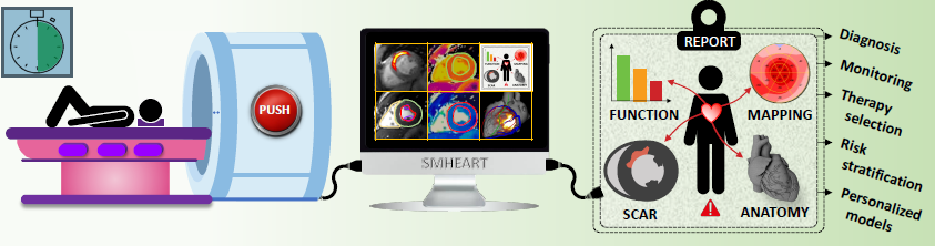
The goal of the SMHEART ERC project is to unleash the full potential of MRI by introducing a fast, one-click, fully automated and comprehensive imaging pipeline applicable to cardiology diagnosis, prognosis and therapy selection.
Current MRI systems are too slow, too complex and require highly trained specialists.
A one-click approach to rapidly collect a single, free-breathing 3D multiparametric volume of the entire heart, thus more comfortable for the patient, with automated extraction of cardiac tissue anatomy, function, and characteristics, using artificial intelligence, will directly improve patient care.
This is not only the condition for wider adoption of MRI in cardiology and the opportunity for better diagnosis, but it also offers the opportunity for improved knowledge of cardiovascular diseases through a multiparametric approach.
The results of this project will pave the way for robust image-based strategies for personalized patient care (diagnosis, risk stratification, therapy selection, monitoring and image-guided interventions).
In concrete terms, what will the funding contribute to?
The funding will allow the recruitment of physicists, mathematicians, computer engineers, and clinicians who will work together at the interface between medical imaging, artificial intelligence, software development, and clinical translation to develop this new imaging solution.
The research project will start in 2023 for 5 years and will first include a pre-clinical exploration phase and then a clinical study that will include cohorts of patients and healthy volunteers.