Multi-modality Platform for Specific Imaging in Cardiology
MUSIC (Multi-modality Platform for Specific Imaging in Cardiology) est une plateforme développée par l’équipe ‘Science des données multimodales’ de l’IHU Liryc ainsi que les équipes Inria Epione et Inria Carmen. L’objectif est d’offrir un large spectre de fonctionnalités et de chaînes de traitements consacrées à la cardiologie, tant pour le diagnostic que pour le guidage thérapeutique. MUSIC est composé de plusieurs modules, comme le logiciel MUSICardio, basé sur le logiciel open source medInria. Il offre :
A propos de l’outil MUSICardio
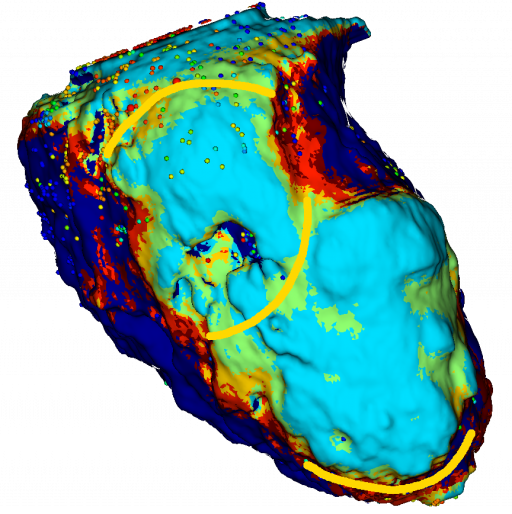
Segmentation, visualisation, filtres, histogramme, reformatage, recalage et outils de gestion de maillages sont proposés et permettent à la communauté d’utiliser un système logiciel ergonomique et plus efficace. De nombreuses modalités d’imagerie sont supportées : MDCT, MRI, PET, maillages VTK, etc. De plus, les modèles générés sont exportables vers les systèmes de cartographie 3D électro-anatomique utilisés par les cardiologues lors des interventions sur les troubles du rythme cardiaque (ablation par cathéter).
MUSICardio propose des algorithmes de traitement d’image à visée diagnostique ou pronostique, des chaînes de traitement destinés au guidage des interventions sur les arythmies atriales et ventriculaires par l’imagerie. Ainsi, MUSICardio a été utilisé pour guider des ablations de tachycardies ventriculaires (TV) dans plus de 300 procédures dans notre centre, et plus de 1500 dans le réseau international MUSIC.
MUSICardio est une plateforme logicielle permettant d’analyser dans un même environnement les données multi-paramétriques issues des diverses équipes de l’IHU (structurelles, mécaniques, hémodynamiques, électriques…), ainsi que d’interfacer avec les plateformes logicielles de simulation type SOFA, CEPS, CARP dans le but de développer des stratégies de modélisation/simulation personnalisées au patient.
Un consortium international de centres spécialisés dans les TV a été créé dans le cadre du projet MUSIC. Il regroupe maintenant plus de 30 centres internationaux. Les centres participants envoient à notre équipe leurs données anonymisées à travers un portail web. Nos manipulateurs radio les traitent en quelques heures et les renvoient au centre partenaire à temps pour l’intervention. Ainsi, des cardiologues du monde entier utilisent chaque jour cette technologie pour guider leurs interventions, visualiser leurs cathéters en temps réel au sein d’un coeur virtuel personnalisé produit à Bordeaux. De plus, ce réseau multicentrique permet de constituer un registre international sur les pathologies structurelles cardiaques incluant données d’imagerie et d’électrophysiologie.
À propos de l’équipe
Notre équipe : le projet MUSIC a été développé par une équipe multidisciplinaire incluant des médecins, chercheurs, informaticiens, et manipulateurs radio.
Contributeurs du projet :
Responsables développement :
- Florent Collot (Développeur)
- Mathilde Merle (Développeuse)
Développeurs et Développeuses :
- Loic Cadour (Développeur)
- Julien Castelneau (Développeur)
- Nordine El Baraka (Développeur)
- Mehdi Juhoor (Développeur)
- Pauline Migerditichan (Développeuse)
- Florian Vichot (Développeur)
Doctorant :
- Jatin Relan (Doctorant)
Coordinateur scientifique :
- Maxime Sermesant (Chercheur)
Coordinateurs cliniques :
- Hubert Cochet (Radiologue) : analyse d’images cardiaques
- Pierre Jais (Cardiologue) : guidage d’interventions
Analyse des données :
- Olivier Baris (Manipulateur radio)
- Ludovic Germain (Manipulateur radio)
- Bruno Soré (Manipulateur radio)
- Jean Michel Thomas (Manipulateur radio)
Chef d’équipe « Imagerie » :
- Bruno Quesson (Chercheur)
Chef d’équipe « Traitement du signal » :
- Rémi Dubois (Chercheur)
Équipe « Modélisation », équipe Inria « Carmen » :
- Nejib Zemzemi (Chercheur)
- Yves Coudière (Chercheur)
Pour nous contacter : ici
Les dernières actualités du projet MUSIC
Découvrez le projet MUSIC vu par Cyril :
Le projet MUSIC détaillé dans l’article « MUSIC: Cardiac Imaging, Modelling and Visualisation Software for Diagnosis and Therapy«
Visionnez la présentation de l’EquipEx Music :
Cliquez ici pour découvrir un article sur MusiCardio, le logiciel de simulation numérique au service de la cardiologie.
Découvrir le reportage « The rhythm of the heart » (vidéo)
Publications scientifiques
- Substrate Modification Using Stereotactic Radioablation to Treat Refractory Ventricular Tachycardia in Patients With Ischemic Cardiomyopathy. Qian PC, Quadros K, Aguilar M, Wei C, Boeck M, Bredfeldt J, Cochet H, Blankstein R, Mak R, Sauer WH, Tedrow UB, Zei PC.JACC Clin Electrophysiol. 2021 Jul 27:S2405-500X(21)00599-5. doi: 10.1016/j.jacep.2021.06.016.
- Pulsed field ablation prevents chronic atrial fibrotic changes and restrictive mechanics after catheter ablation for atrial fibrillation. Nakatani Y, Sridi-Cheniti S, Cheniti G, Ramirez FD, Goujeau C, André C, Nakashima T, Eggert C, Schneider C, Viswanathan R, Krisai P, Takagi T, Kamakura T, Vlachos K, Derval N, Duchateau J, Pambrun T, Chauvel R, Reddy VY, Montaudon M, Laurent F, Sacher F, Hocini M, Haïssaguerre M, Jaïs P, Cochet H.Europace. 2021 Jul 8:euab155. doi: 10.1093/europace/euab155.
- Cardiac Magnetic Resonance Imaging and Ventricular Tachycardias Involving the Sinuses of Valsalva in Patients With Nonischemic Cardiomyopathy. Ghannam M, Liang JJ, Attili A, Cochet H, Jais P, Latchamsetty R, Jongnarangsin K, Morady F, Bogun F.JACC Clin Electrophysiol. 2021 Jun 22:S2405-500X(21)00282-6. doi: 10.1016/j.jacep.2021.03.017.
- Role of endocardial ablation in eliminating an epicardial arrhythmogenic substrate in patients with Brugada syndrome. Kamakura T, Cochet H, Juhoor M, Nakatani Y, Ramirez FD, André C, Nakashima T, Krisai P, Takagi T, Tixier R, Chauvel R, Cheniti G, Duchateau J, Pambrun T, Derval N, Kusano K, Sacher F, Jaïs P, Haïssaguerre M, Hocini M.Heart Rhythm. 2021 Oct;18(10):1673-1681. doi: 10.1016/j.hrthm.2021.06.1188.
- Deep learning formulation of electrocardiographic imaging integrating image and signal information with data-driven regularization. Bacoyannis T, Ly B, Cedilnik N, Cochet H, Sermesant M.Europace. 2021 Mar 4;23(23 Suppl 1):i55-i62. doi: 10.1093/europace/euaa391.
- The value of cardiac magnetic resonance imaging and programmed ventricular stimulation in patients with ventricular noncompaction and ventricular arrhythmias. Gunda S, Ghannam M, Liang JJ, Attili A, Sharaf Dabbagh G, Cochet H, Lathkar-Pradhan S, Latchamsetty R, Jongnarangsin K, Morady F, Bogun F.J Cardiovasc Electrophysiol. 2021 Mar;32(3):745-754. doi: 10.1111/jce.14884.
- Impact of Intramural Scar on Mapping and Ablation of Premature Ventricular Complexes. Ghannam M, Liang JJ, Dabbagh GS, Siontis KC, Attili A, Cochet H, Jais P, Juhoor M, Latchamsetty R, Jongnarangsin K, Morady F, Bogun F.JACC Clin Electrophysiol. 2021 Jun;7(6):733-741. doi: 10.1016/j.jacep.2020.11.004.
- Magnetic Resonance Mapping of Catheter Ablation Lesions After Post-Infarction Ventricular Tachycardia Ablation. Dabbagh GS, Ghannam M, Siontis KC, Attili A, Cochet H, Jais P, Eng MJ, Latchamsetty R, Jongnarangsin K, Morady F, Bogun F.JACC Cardiovasc Imaging. 2021 Mar;14(3):588-598. doi: 10.1016/j.jcmg.2020.08.041.
- Clinical significance of myocardial scar in patients with frequent premature ventricular complexes undergoing catheter ablation. Ghannam M, Yokokawa M, Liang JJ, Cochet H, Jais P, Dabagh GS, Latchamsetty R, Jongnarangsin K, Morady F, Bogun F.Heart Rhythm. 2021 Jan;18(1):20-26. doi: 10.1016/j.hrthm.2020.07.030.
- Ventricular tachycardia in a patient with repaired d-transposition of the great arteries. Krisai P, Vlachos K, Tafer N, Cochet H, Iriart X, Sacher F.HeartRhythm Case Rep. 2020 Oct 17;7(1):26-29. doi: 10.1016/j.hrcr.2020.10.006.
- Double ventricular tachycardias associated with an anatomical isthmus identified by a computed tomography-derived channel. Takigawa M, Martin R, Kitamura T, Cochet H, Jais P, Sacher F.J Cardiovasc Electrophysiol. 2020 Nov;31(11):3061-3063. doi: 10.1111/jce.14735.
- Risk stratification in patients with nonischemic cardiomyopathy and ventricular arrhythmias based on quantification of intramural delayed enhancement on cardiac magnetic resonance imaging. Ghannam M, Siontis KC, Cochet H, Jais P, Juhoor M, Attili A, Sharaf-Dabbagh G, Latchamsetty R, Jongnarangsin K, Morady F, Bogun F.J Cardiovasc Electrophysiol. 2020 Jul;31(7):1762-1769. doi: 10.1111/jce.14514.
- Stepwise Approach for Ventricular Tachycardia Ablation in Patients With Predominantly Intramural Scar. Ghannam M, Siontis KC, Kim HM, Cochet H, Jais P, Juhoor M, Latchamsetty R, Jongnarangsin K, Attili A, Sharaf Dabbagh G, Yokokawa M, Morady F, Bogun F.JACC Clin Electrophysiol. 2020 Apr;6(4):448-460. doi: 10.1016/j.jacep.2019.11.020.
- Value of mapping and ablation of ventricular tachycardia targets within the coronary venous system in patients with nonischemic cardiomyopathy. Ghannam M, Siontis KC, Cochet H, Jais P, Eng MJ, Latchamsetty R, Jongnarangsin K, Dabbagh GS, Yokokawa M, Morady F, Bogun F.Heart Rhythm. 2020 Apr;17(4):520-526. doi: 10.1016/j.hrthm.2020.01.010.
- Image-guided ablation of scar-related ventricular tachycardia: towards a shorter and more predictable procedure. Berte B, Cochet H, Dang L, Mahida S, Moccetti F, Hilfiker G, Bondietti J, Ruschitzka F, Jaïs P, Scharf C, Kobza R.J Interv Card Electrophysiol. 2020 Dec;59(3):535-544. doi: 10.1007/s10840-019-00686-w.
Risk stratification in patients with frequent premature ventricular complexes in the absence of known heart disease. Ghannam M, Siontis KC, Kim MH, Cochet H, Jais P, Eng MJ, Attili A, Sharaf-Dabbagh G, Latchamsetty R, Jongnarangsin K, Morady F, Bogun F.Heart Rhythm. 2020 Mar;17(3):423-430. doi: 10.1016/j.hrthm.2019.09.027.
- A Spatial Adaptation of the Time Delay Neural Network for Solving ECGI Inverse Problem. Amel Karoui, Mostafa Bendahmane, Nejib Zemzemi, Yves Coudière; Valéry Ozenne; Edward Vigmond; Nejib Zemzemi. 10th International Symposium Functional Imaging and Modeling of the Heart, 11504, Springer, pp.94-102, 2019, Lecture Notes in Computer Science, 978-3-030-21949-9. ⟨10.1007/978-3-030-21949-9_11⟩
- Berte et al.Image-guided ablation of scar-related ventricular tachycardia: towards a shorter and more predictable procedure. Journal of Interventional Cardiac Electrophysiology – 2019 Dec.
- Direct Mapping from Body Surface Potentials to Cardiac Activation Maps Using Neural Networks. Amel Karoui, Mostafa Bendahmane, Nejib Zemzemi. CinC 2019 – 46th Computing in Cardiology Conference, Sep 2019,Singapour, Singapore
- Space rescaling in the MFS method improves the ECGI reconstruction. Pauline Migerditichan, Mark Potse, Nejib Zemzemi. CinC 2019 – Computing in Cardiology 2019, Sep 2019, Singapour, Singapore
- Three-dimensional image integration guidance for cryoballoon pulmonary vein isolation procedures.
Bourier F, Vlachos K, Lam A, Martin CA, Takigawa M, Kitamura T, Massoullié G, Cheniti G, Frontera A, Duchateau J, Pambrun T, Klotz N, Derval N, Denis A, Hocini M, Haïssaguerre M, Cochet H, Jaïs P, Sacher F.J Cardiovasc Electrophysiol. 2019 Dec;30(12):2790-2796. doi: 10.1111/jce.14249. - Post-Myocardial Infarction Scar With Fat Deposition Shows Specific Electrophysiological Properties and Worse Outcome After Ventricular Tachycardia Ablation.
Cheniti G, Sridi S, Sacher F, Chaumeil A, Pillois X, Takigawa M, Frontera A, Vlachos K, Martin CA, Teijeira E, Kitamura T, Lam A, Bourier F, Puyo S, Duchateau J, Denis A, Pambrun T, Chauvel R, Derval N, Laurent F, Montaudon M, Hocini M, Haissaguerre M, Jais P, Cochet H.J Am Heart Assoc. 2019 Aug 6;8(15):e012482. doi: 10.1161/JAHA.119.012482. - Are wall thickness channels defined by computed tomography predictive of isthmuses of postinfarction ventricular tachycardia?
Takigawa M, Duchateau J, Sacher F, Martin R, Vlachos K, Kitamura T, Sermesant M, Cedilnik N, Cheniti G, Frontera A, Thompson N, Martin C, Massoullie G, Bourier F, Lam A, Wolf M, Escande W, André C, Pambrun T, Denis A, Derval N, Hocini M, Haissaguerre M, Cochet H, Jaïs P.Heart Rhythm. 2019 Nov;16(11):1661-1668. doi: 10.1016/j.hrthm.2019.06.012. - Focal scar and diffuse myocardial fibrosis are independent imaging markers in repaired tetralogy of Fallot.
Cochet H, Iriart X, Allain-Nicolaï A, Camaioni C, Sridi S, Nivet H, Fournier E, Dinet ML, Jalal Z, Laurent F, Montaudon M, Thambo JB.Eur Heart J Cardiovasc Imaging. 2019 Sep 1;20(9):990-1003. doi: 10.1093/ehjci/jez068. - Relationship between atrial scar on cardiac magnetic resonance and pulmonary vein reconnection after catheter ablation for paroxysmal atrial fibrillation.
Jefairi NA, Camaioni C, Sridi S, Cheniti G, Takigawa M, Nivet H, Denis A, Derval N, Mathilde Merle, Laurent F, Montaudon M, Sacher F, Hocini M, Haissaguerre M, Jais P, Cochet H.J Cardiovasc Electrophysiol. 2019 May;30(5):727-740. doi: 10.1111/jce.13908. - Takigawa et al.Detailed comparison between the wall thickness and voltages in chronic myocardial infarction. Journal of Cardiovascular Electrophysiology – 2019 Feb;30(2):195-204.
- Cabrera Lozoya et al.Model-Based Feature Augmentation for Cardiac Ablation Target Learning From Images.IEEE Transactions in Biomedical Engineering – 2019 Jan;66(1):30-40.
- Evaluation of fifteen algorithms for the resolution of the electrocardiography imaging inverse problem using ex-vivo and in-silico data. Amel Karoui, Laura Bear, Pauline Migerditichan, Nejib Zemzemi. Frontiers in Physiology, Frontiers, 2018, Electrocardiographic Imaging, 9, pp.1708
- Fast personalized electrophysiological models from computed tomography images for ventricular tachycardia ablation planning.
Cedilnik N, Duchateau J, Dubois R, Sacher F, Jaïs P, Cochet H, Sermesant M.Europace. 2018 Nov 1;20(suppl_3):iii94-iii101. doi: 10.1093/europace/euy228. - Patient-specific simulations predict efficacy of ablation of interatrial connections for treatment of persistent atrial fibrillation.
Roney CH, Williams SE, Cochet H, Mukherjee RK, O’Neill L, Sim I, Whitaker J, Razeghi O, Klein GJ, Vigmond EJ, O’Neill M, Niederer SA.Europace. 2018 Nov 1;20(suppl_3):iii55-iii68. doi: 10.1093/europace/euy232. - The Heart Recording Conditions Impact the Assessment of the Electrocardiography Imaging Inverse Solution. Amel Karoui, Laura Bear, Pauline Migerditichan, Mostafa Bendahmane, Nejib Zemzemi.CinC 2018 – 45th Computing in Cardiology Conference, Sep 2018, Maastricht, Netherlands
- Teijeira‐Fernandez et al.Influence of contact force on voltage mapping: A combined magnetic resonance imaging and electroanatomic mapping study in patients with tetralogy of fallot. Heart Rhythm – 2018 Aug;15(8):1198-1205.
- Ghannam et al.Correlation between computer tomography‐derived scar topography and critical ablation sites in postinfarction ventricular tachycardia. Journal of Cardiovascular Electrophysiology – 2018 Mar;29(3):438‐445.
- Wolf et al.Long-term outcome of substrate modification in ablation of post-myocardial infarction ventricular tachycardia Circulation: Arrhythmia and Electrophysiology – 2018 Feb;11(2):e005635.
- Cochet et al.Relationship between fibrosis detected on late gadolinium-enhanced cardiac magnetic resonance and re-entrant activity assessed with electrocardiographic imaging in human persistent atrial fibrillation. JACC: Clinical Electrophysiology – 2018 Jan;4(1):17-29.
- Mahida et al.Cardiac imaging in patients with ventricular tachycardia. Circulation – 2017 Dec 19;136(25):2491-2507.
- Pierre Jaïs.CT scan isthmuses as an imaging target for VT ablation. International Symposium on Ventricular Arrhythmias – 2017.
- Cedilnik et al.VT scan: Towards an efficient pipeline from computed tomography images to ventricular tachycardia ablation. Functional Imaging and Modelling of the Heart (FIMH) – 2017, Lecture Notes in Computer Science, vol 10263.
- Thompson et al..Catheter ablation for ventricular tachycardia in patients with nonischemic cardiomyopathy. Cardiac Electrophysiology Clinics – 2017 Mar;9(1):47-54.
- Yamashita S, et al. Myocardial wall thinning predicts transmural substrate in patients with scar-related ventricular tachycardia. Heart Rhythm. 2017 Feb;14(2):155-163.
- Cabrera-Lozoya R, et al. Image-Based Biophysical Simulation of Intracardiac Abnormal Ventricular Electrograms. IEEE Trans Biomed Eng. 2017 Jul;64(7):1446-1454.
- Roney CH, et al. Modelling methodology of atrial fibrosis affects rotor dynamics and electrograms. Europace. 2016 Dec;18(suppl 4):iv146-iv155.
- Yamashita S, et al. Impact of New Technologies and Approaches for Post-Myocardial Infarction Ventricular Tachycardia Ablation During Long-Term Follow-Up. Circ Arrhythm Electrophysiol. 2016 Jul;9(7).
- Zahid S, et al. Patient-derived models link re-entrant driver localization in atrial fibrillation to fibrosis spatial pattern. Cardiovasc Res. 2016 Jun 1;110(3):443-54.
- Vigmond E, et al. Percolation as a mechanism to explain atrial fractionated electrograms and reentry in a fibrosis model based on imaging data. Heart Rhythm. 2016 Jul;13(7):1536-43.
- Yamashita S, et al. Image Integration to Guide Catheter Ablation in Scar-Related Ventricular Tachycardia. J Cardiovasc Electrophysiol. 2016 Jun;27(6):699-708.
- Haissaguerre M, et al. Intermittent drivers anchoring to structural heterogeneities as a major pathophysiological mechanism of human persistent atrial fibrillation. J Physiol. 2016 May 1;594(9):2387-98.
- Berte B, et al. Irrigated Needle Ablation Creates Larger and More Transmural Ventricular Lesions Compared With Standard Unipolar Ablation in an Ovine Model. Circ Arrhythm Electrophysiol. 2015 Dec;8(6):1498-506.
- Berte B, et al. Characterization of the Left-Sided Substrate in Arrhythmogenic Right Ventricular Cardiomyopathy. Circ Arrhythm Electrophysiol. 2015 Aug 26. pii: CIRCEP.115.003213. [Epub ahead of print]
- Berte B, et al. Epicardial only mapping and ablation of ventricular tachycardia: a case series. Europace. 2015 Apr 2. pii: euv072. [Epub ahead of print]
- Cochet H, Mouries A, Nivet H, Sacher F, Derval N, Denis A, Merle M, Relan J, Hocini M, Haïssaguerre M, Laurent F, Montaudon M, Jaïs P. Age, atrial fibrillation, and structural heart disease are the main determinants of left atrial fibrosis detected by delayed-enhanced magnetic resonance imaging in a general cardiology population. J Cardiovasc Electrophysiol. 2015 May;26(5):484-92.
- Yamashita S, et al. Role of high-resolution image integration to visualize left phrenic nerve and coronary arteries during epicardial ventricular tachycardia ablation. Circ Arrhythm Electrophysiol. 2015 Apr;8(2):371-80.
- Cochet H, et al. Automated Quantification of Right Ventricular Fat at Contrast-enhanced Cardiac Multidetector CT in Arrhythmogenic Right Ventricular Cardiomyopathy. Radiology. 2015 Jan 5:141140. [Epub ahead of print]
- Berte B, et al. Postmyocarditis ventricular tachycardia in patients with epicardial-only scar: a specific entity requiring a specific approach. J Cardiovasc Electrophysiol. 2015 Jan;26(1):42-50.
- Labarthe S, et al. A bilayer model of human atria: mathematical background, construction, and assessment. Europace. 2014 Nov;16 Suppl 4:iv21-iv29.
- Komatsu Y, et al. Relationship between MDCT-imaged myocardial fat and ventricular tachycardia substrate in arrhythmogenic right ventricular cardiomyopathy. J Am Heart Assoc. 2014 Aug 7;3(4).
- Cochet H, et al. Atrial structure and function 5 years after successful ablation for persistent atrial fibrillation: an MRI study. J Cardiovasc Electrophysiol. 2014 Jul;25(7):671-9.
- Cochet H, et al. Cardiac arrythmias: multimodal assessment integrating body surface ECG mapping into cardiac imaging. Radiology. 2014 Apr;271(1):239-47.
- Vigmond E, et al. A bilayer representation of the human atria. Conf Proc IEEE Eng Med Biol Soc. 2013;2013:1530-3.
- Komatsu Y, et al. Multimodality imaging to improve the safety and efficacy of epicardial ablation of scar-related ventricular tachycardia. J Cardiovasc Electrophysiol. 2013 Dec;24(12):1426-7.
- Jadidi AS, et al. Inverse relationship between fractionated electrograms and atrial fibrosis in persistent atrial fibrillation: combined magnetic resonance imaging and high-density mapping. J Am Coll Cardiol 2013;62:802-12.
- Komatsu Y, et al. Regional myocardial wall thinning at multidetector computed tomography correlates to arrhythmogenic substrate in postinfarction ventricular tachycardia: assessment of structural and electrical substrate. Circ Arrhythm Electrophysiol 2013;6:342-50.
- Hocini M, et al. Noninvasive electrocardiomapping facilitates previously failed ablation of right appendage diverticulum associated life-threatening accessory pathway. J Cardiovasc Electrophysiol 2013;24:583-5.
- Cochet H, et al. Integration of merged delayed-enhanced magnetic resonance Imaging and multidetector computed tomography for the guidance of ventricular tachycardia ablation: a pilot study.J Cardiovasc Electrophysiol 2013;24:419-26.